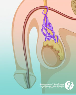
Port-A-Cath is a type of central venous catheter for patients who require long-term venous access to protect the patient's blood vessels from frequent blood draws and chemotherapy drugs that can cause damage to the blood vessels.
PortaCath consists of a small disc implanted under the skin in the chest. The disc has a small port that connects to a tube that extends into a large vein. A special needle is used to access the port and deliver treatment or draw blood samples.
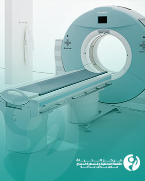
Interventional radiological placement of the PortaCath ensures precise guidance for placing the PortaCath in the exact location and minimizes the risk of infection.
You can contact the Al Hayat Center for Interventional Radiology and Neurointervention for non-surgical treatment through the phone number 07744700048
Reasons for installing Portacath:
Portacath is primarily inserted to provide long-term central venous access. This is particularly useful in the following situations:
• Long-term administration of medication (such as chemotherapy or antibiotics).
• Difficult or impossible venous access using traditional techniques.
• Drawing blood samples in patients who require frequent blood tests.
• Providing parenteral nutrition.
PortaCath Privileges:
PortaCath offers many benefits, including:
• It reduces the need for frequent vein pokes and minimizes the risk of phlebitis.
• It provides easy access to long-term treatments without having to find veins every time.
• Portacath can last for up to several years.
• Since it is located under the skin, PortaCath requires little care.
• PortaCath is water-resistant, which facilitates normal daily handling.
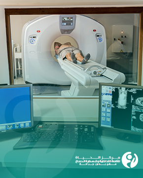
Steps of Portacath insertion and the role of interventional radiology in Portacath placement:
The Port-A-Cath fitted using interventional radiology is securely fixed under the skin, and can be used for a relatively long time without needing to be changed. Here is an overview of how this interventional radiological procedure is performed:
1. Medical assessment: The patient's health condition and the need for PortaCath are assessed based on the medical need.
2. Patient preparation: The patient is instructed to fast for several hours before the procedure by medical guidelines.
3. Local anesthesia: The patient is given a local anesthetic to numb the area where the insertion will be performed.
4. Sterilizing the area: The area selected for the installation of the Portacath is sterilized with sterilizers to reduce the risk of infection.
5. Catheter guidance for PortaCath insertion: Using interventional radiology, the doctor guides the PortaCath to the target vein and checks its precise position. First, the radiologist inserts a catheter, a small tube into the vein, into the neck. Next, a small pocket is made under the skin of the chest below the collarbone so that the PortaCath fits into the pocket. The tube attached to the PortaCath is then placed under the skin of the chest so that it enters the neck vein, all under X-ray guidance.
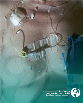
6. Portacath fixation: The port is permanently fixed under the skin, and the wound is carefully closed.
7. Functional testing: The Portacath is frequently tested to check that there is not too much bleeding and that it is functioning well and can receive treatments effectively.
8. Post-procedure follow-up: The patient is followed up to ensure no complications and receives instructions on wound care and how to proceed with treatment.
If you need frequent intravenous medications or frequent blood draws, PortaCath can eliminate the need for repeated vein punctures. Once placed under the skin, it remains discreet and unobtrusive. Contact the Al Hayat Center for Interventional Radiology and Neurointervention at Warith International Cancer Institute to book your consultation with the best interventional radiologists.